


2
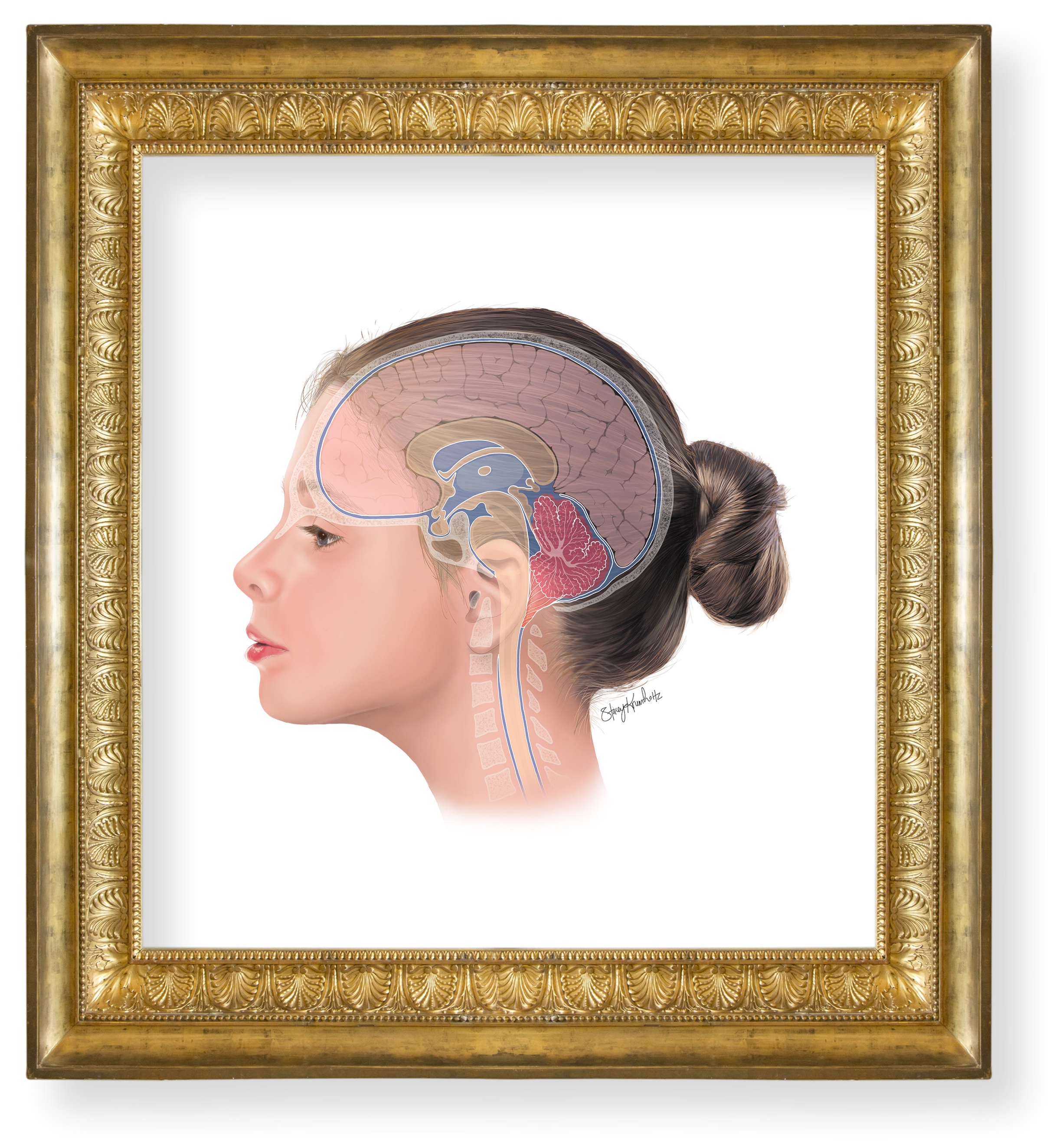
Chiari Malformation
1
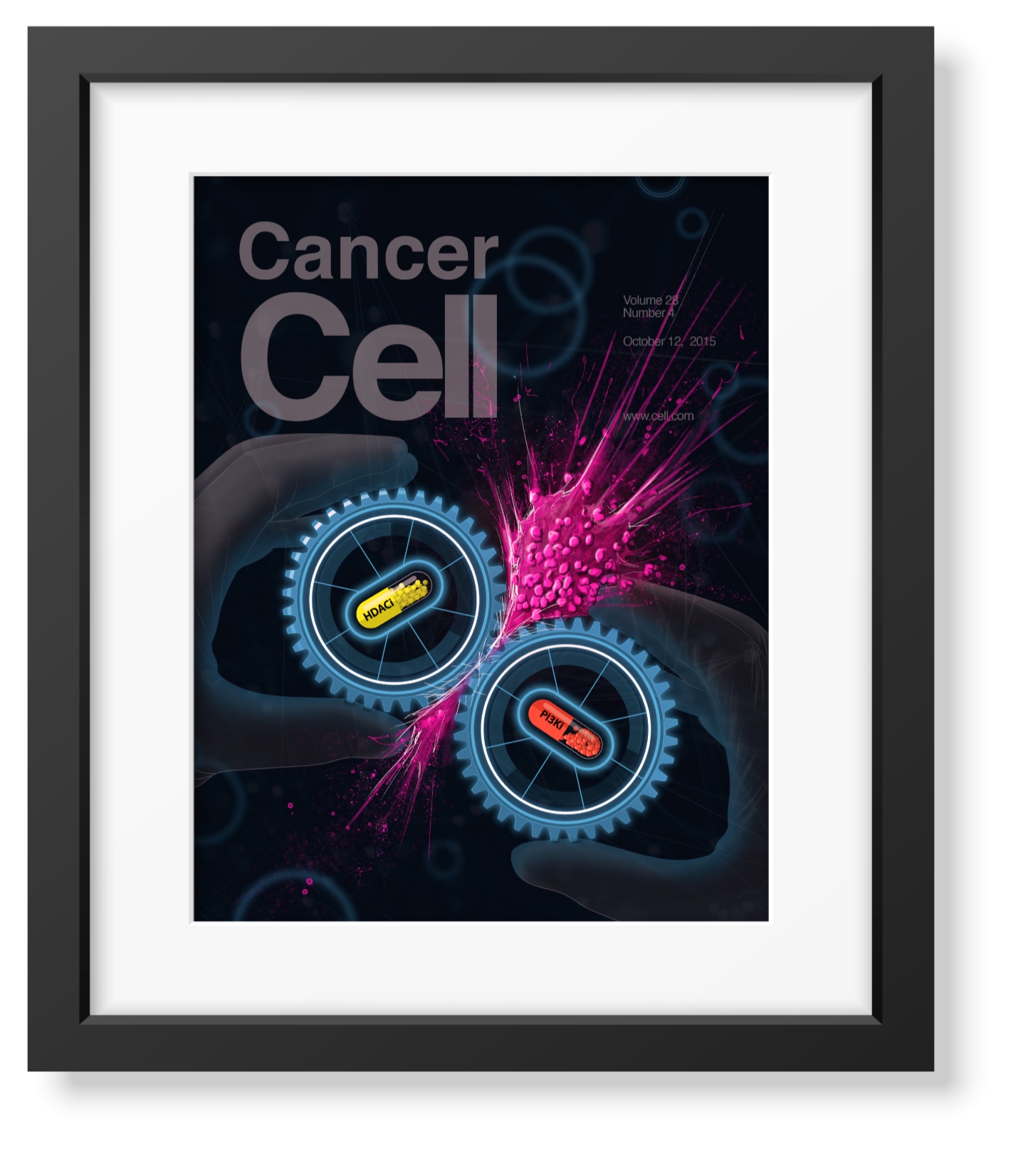
Drug Synergy
3
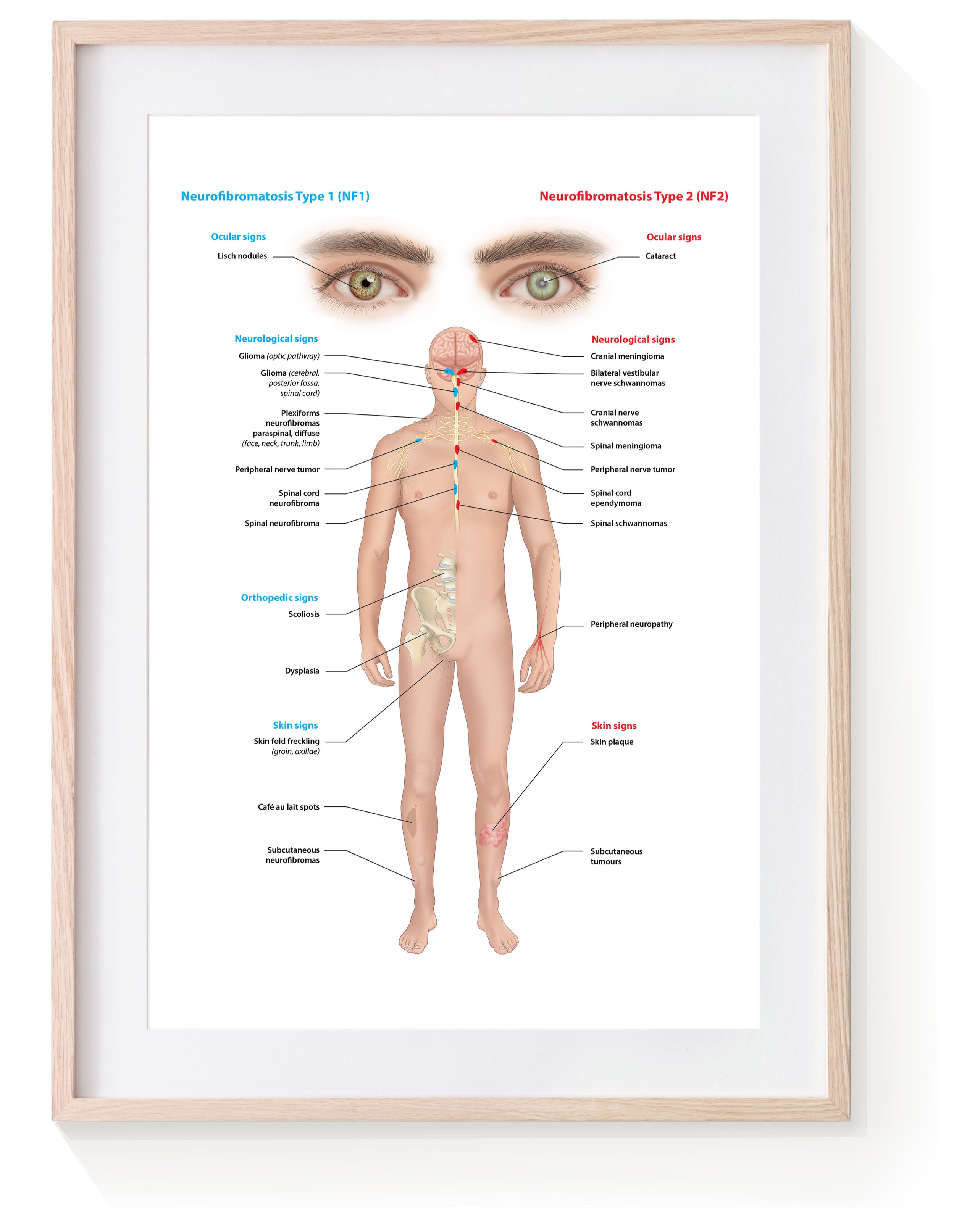
NF1 vs NF2
1
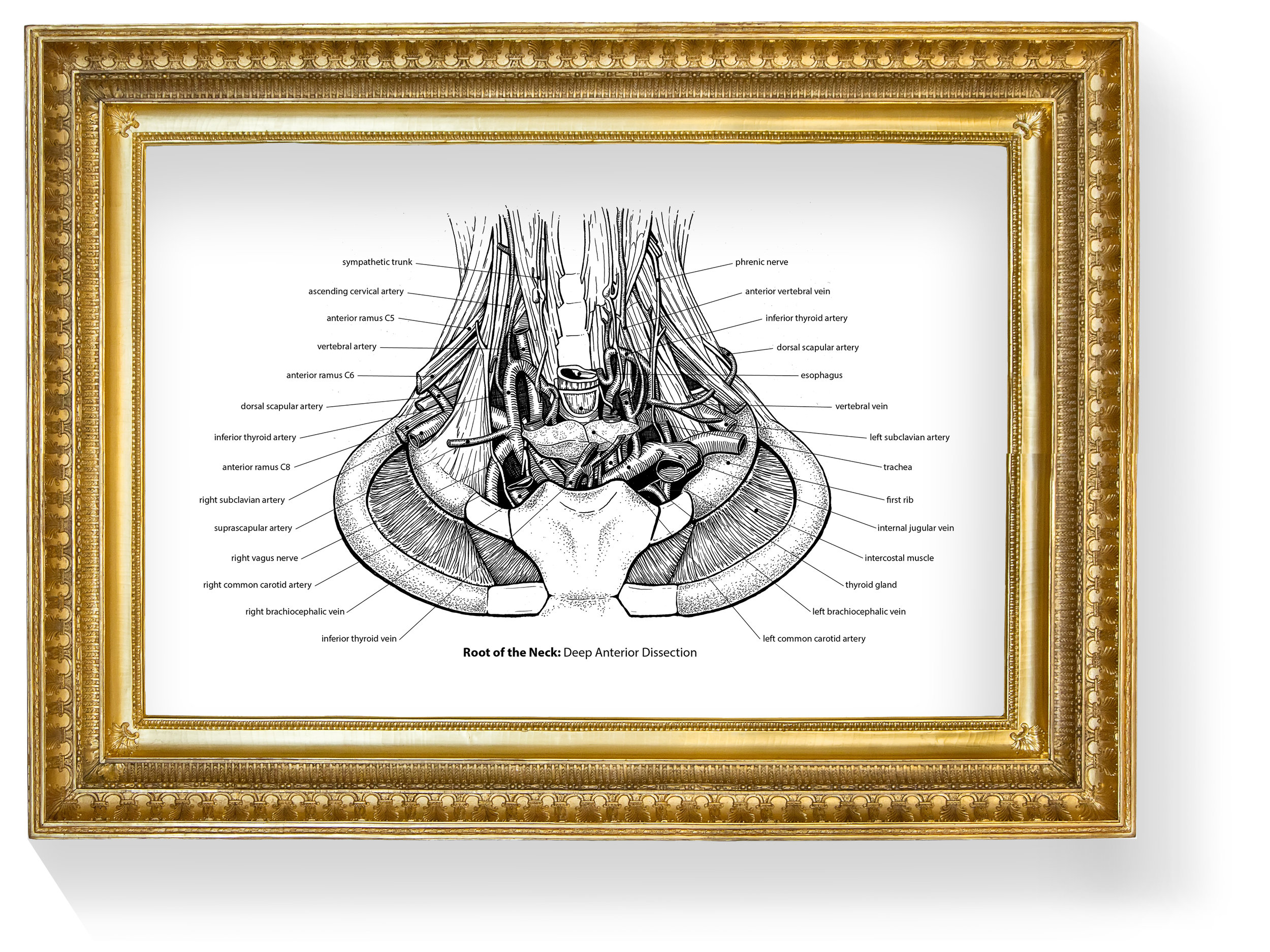
Root of the Neck
2
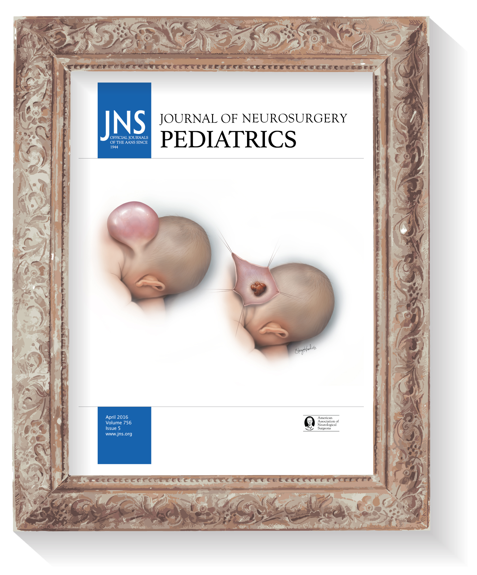
Occipital Encephalocele
2
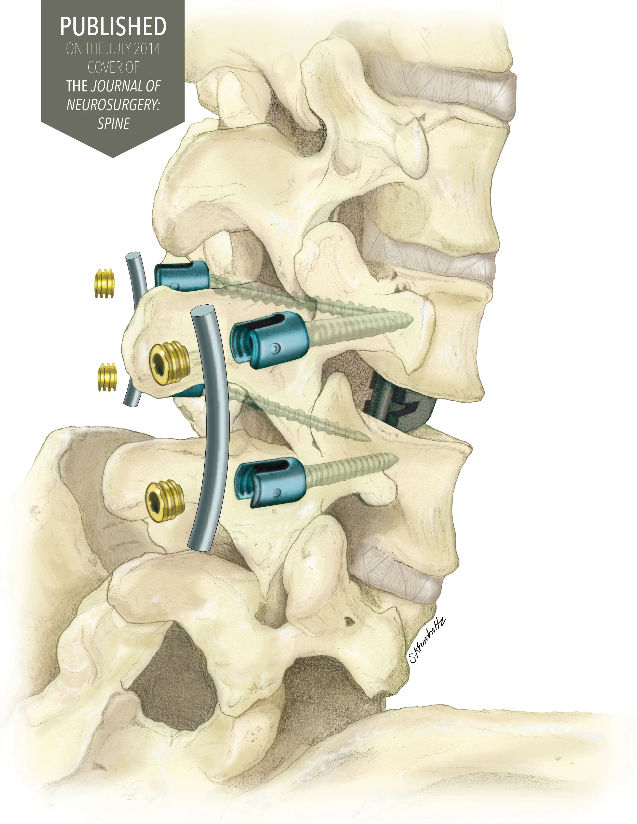
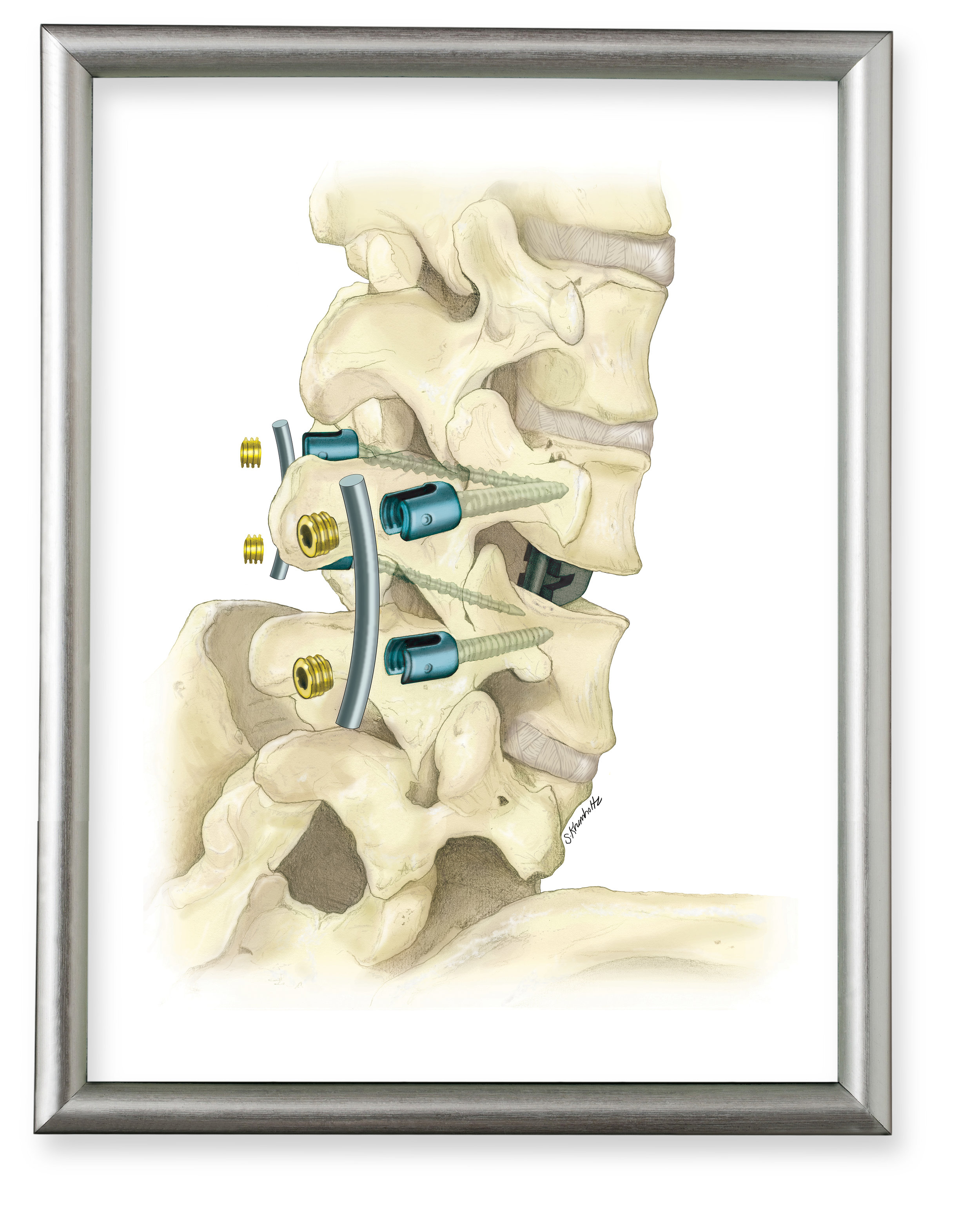
Lumbar Pedicle Fusion
5
Posterior Fossa Tumour

5
Craniosynostosis
1
Tumor Progression
2
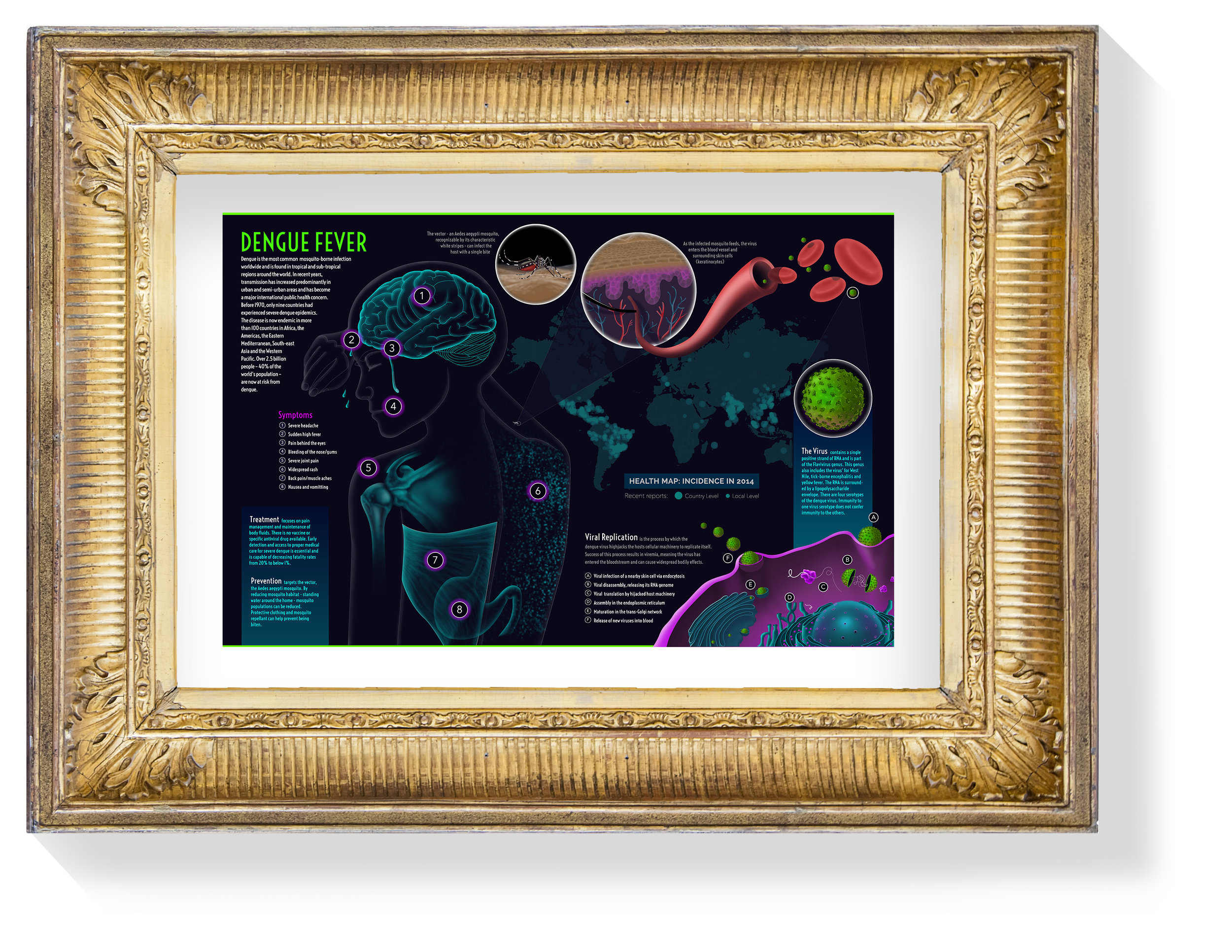
Dengue Fever
1
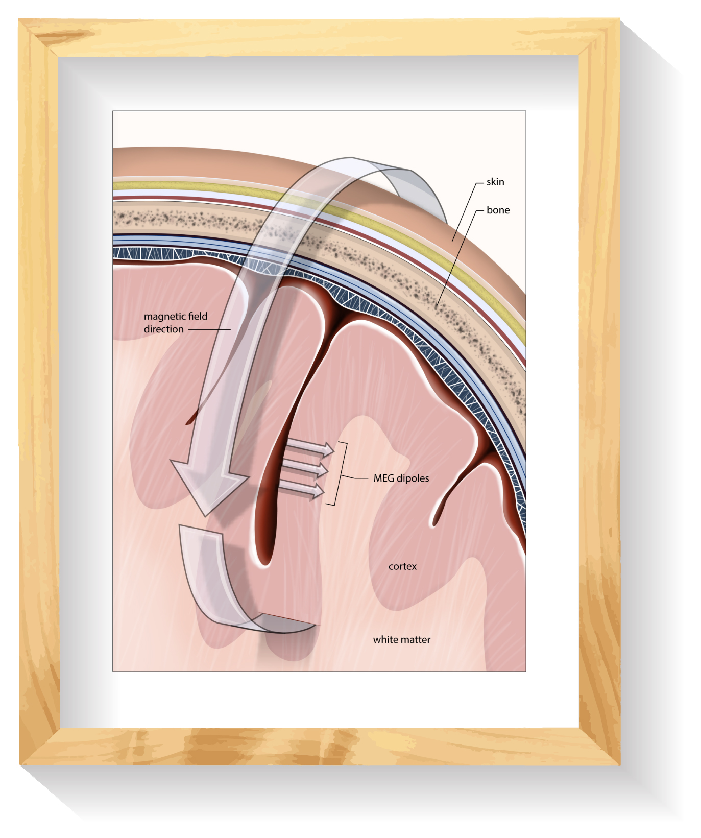
MEG
1
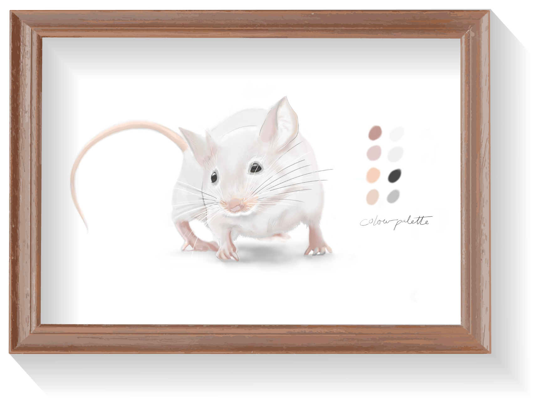
Mouse Figures
11
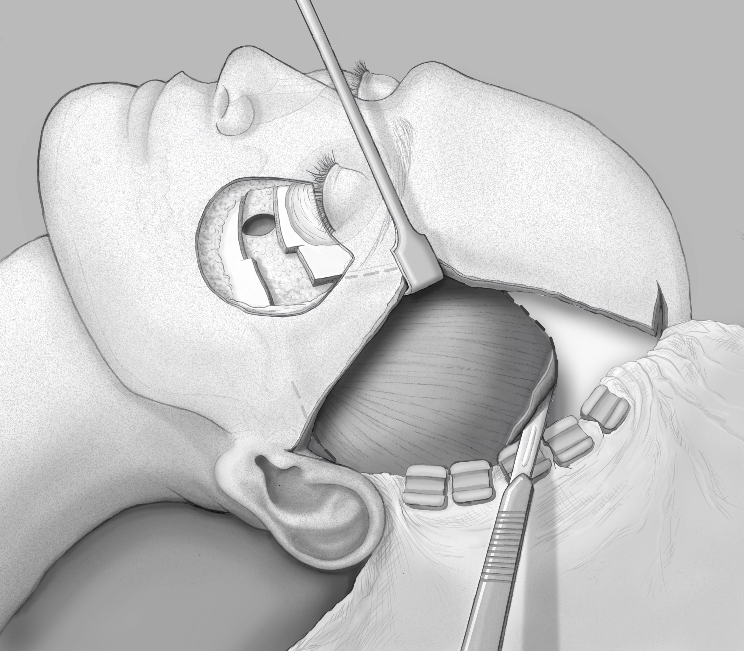
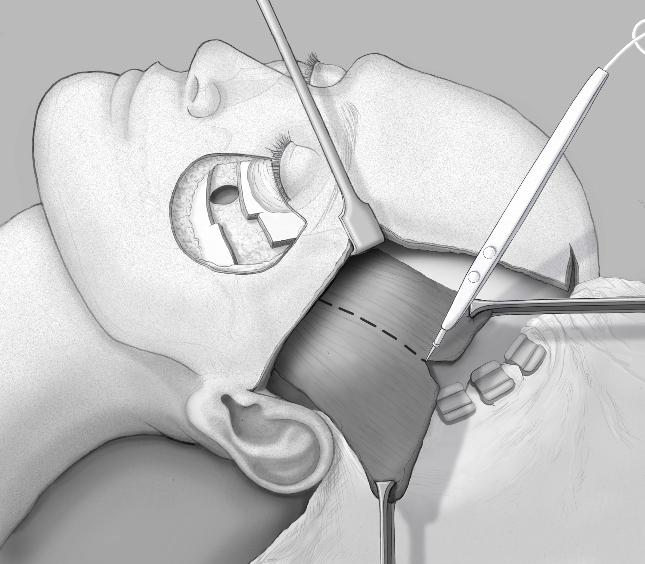
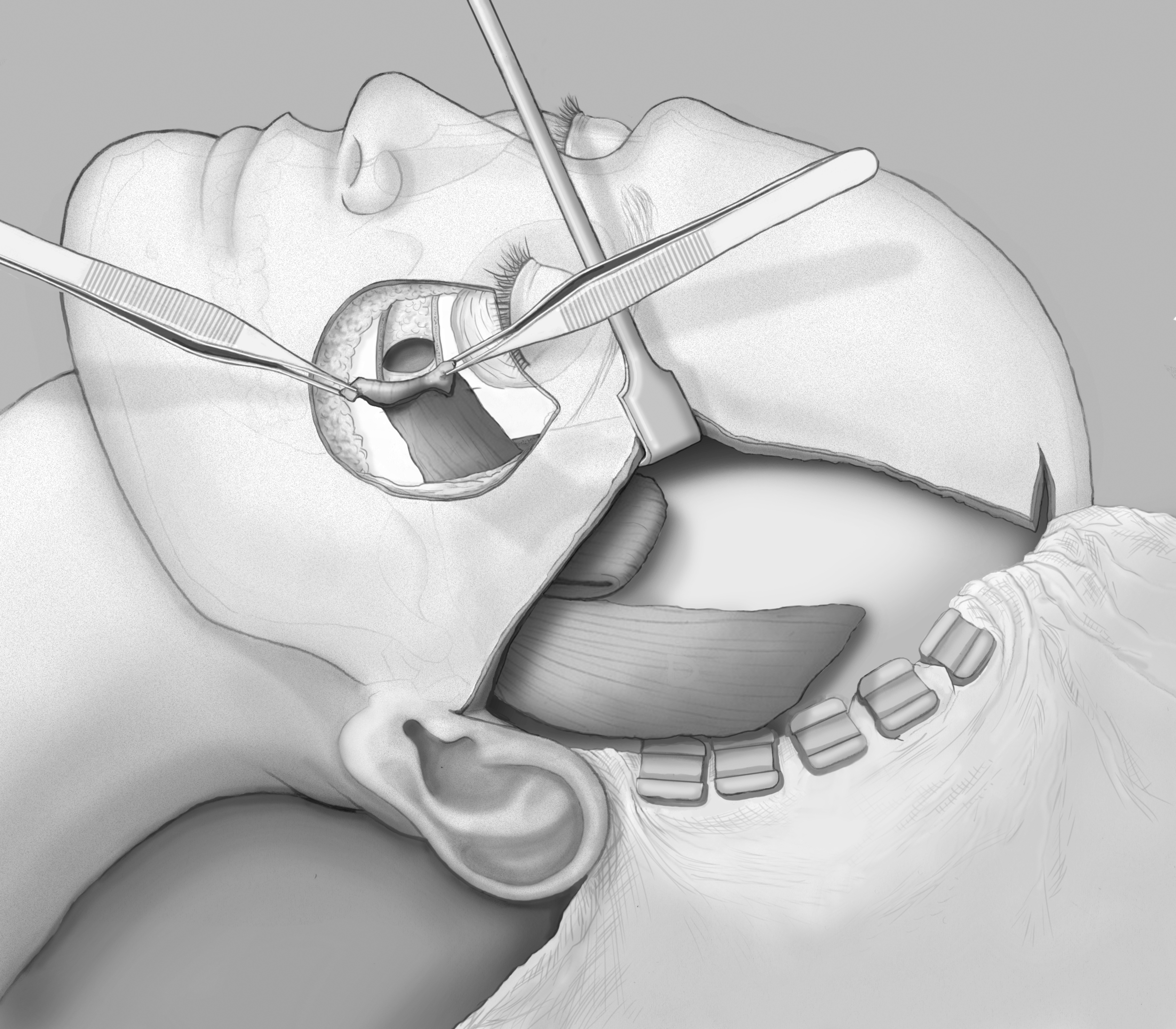
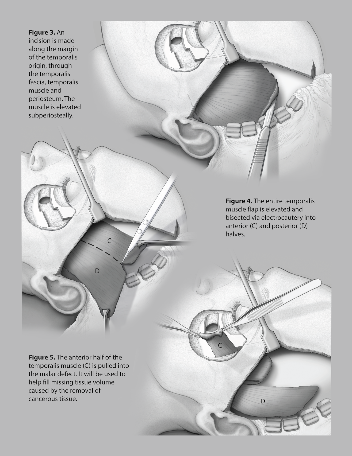
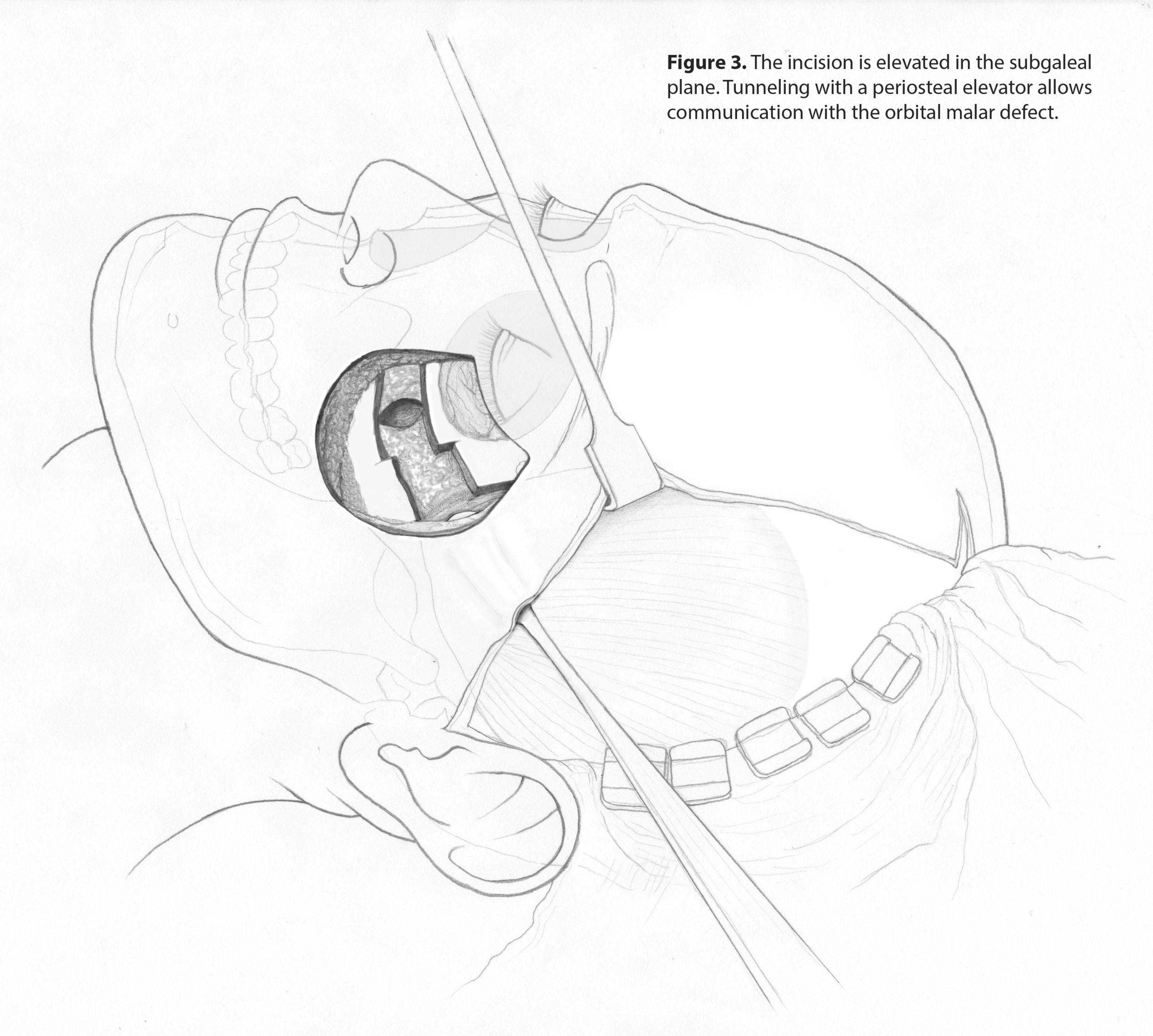
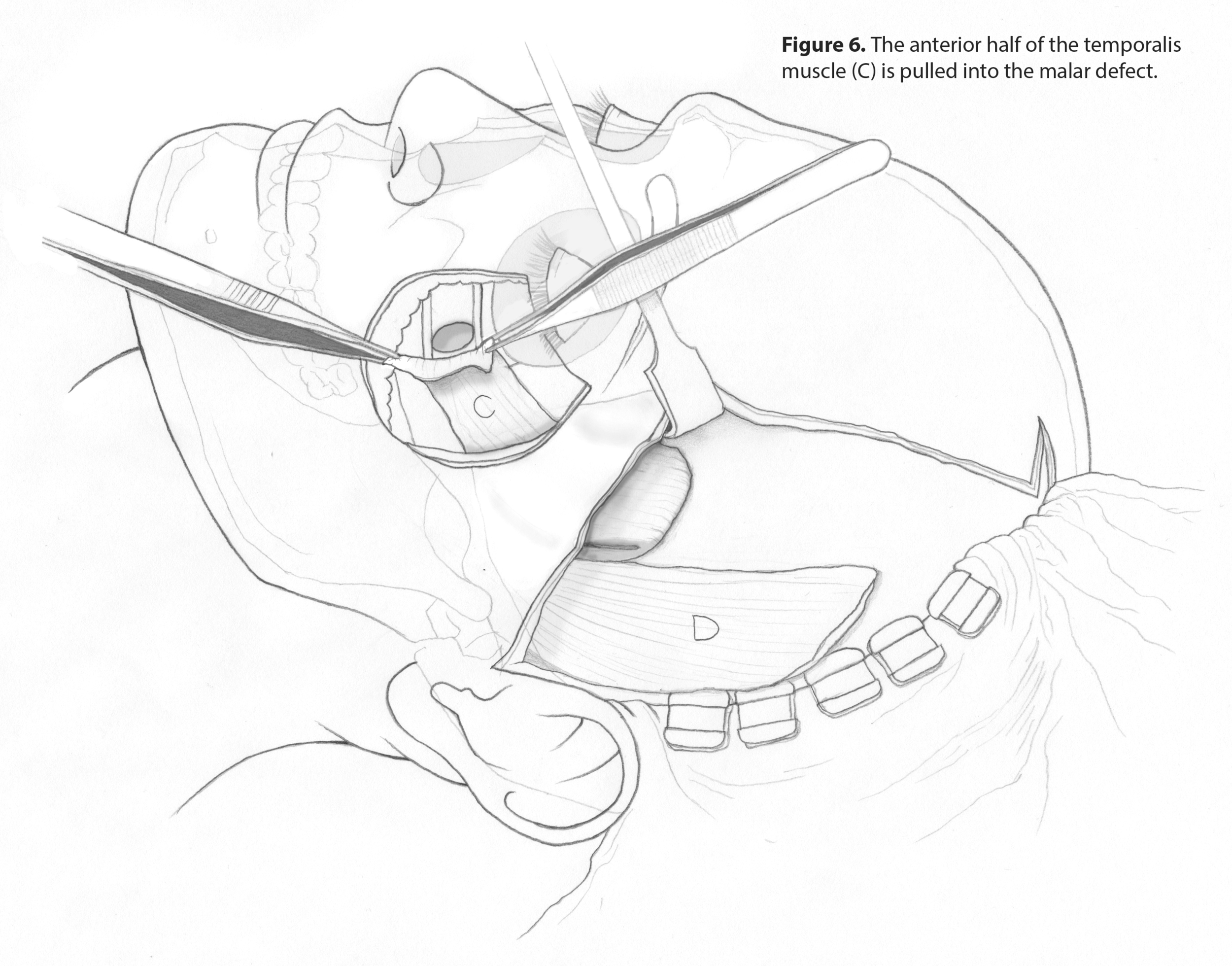
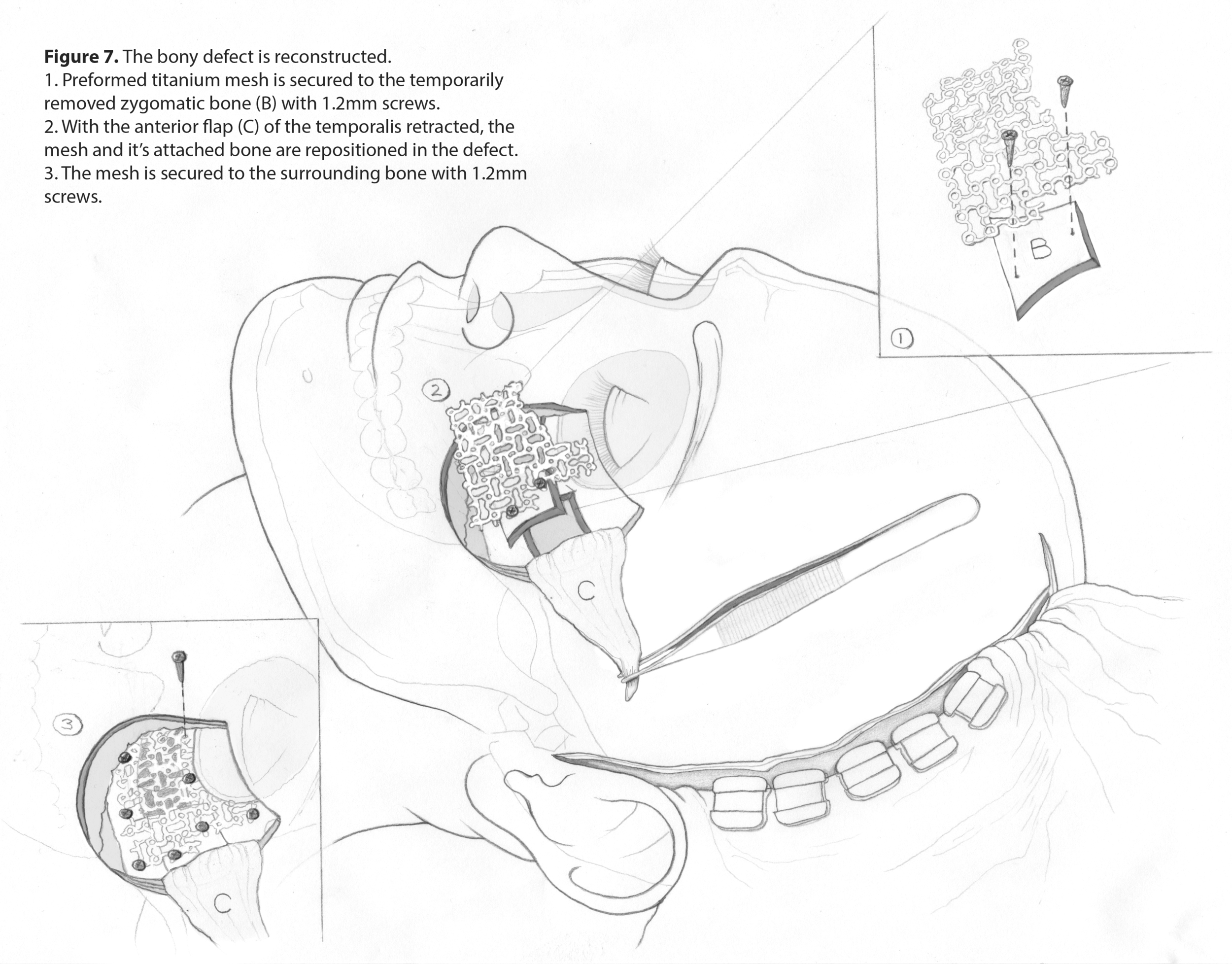
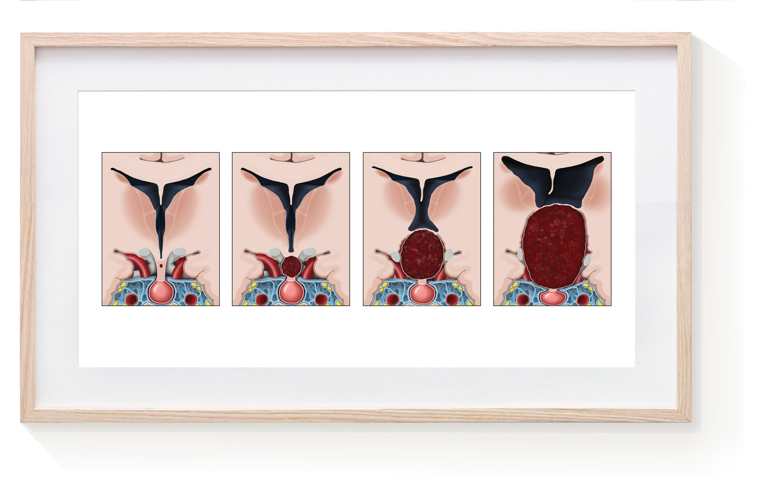
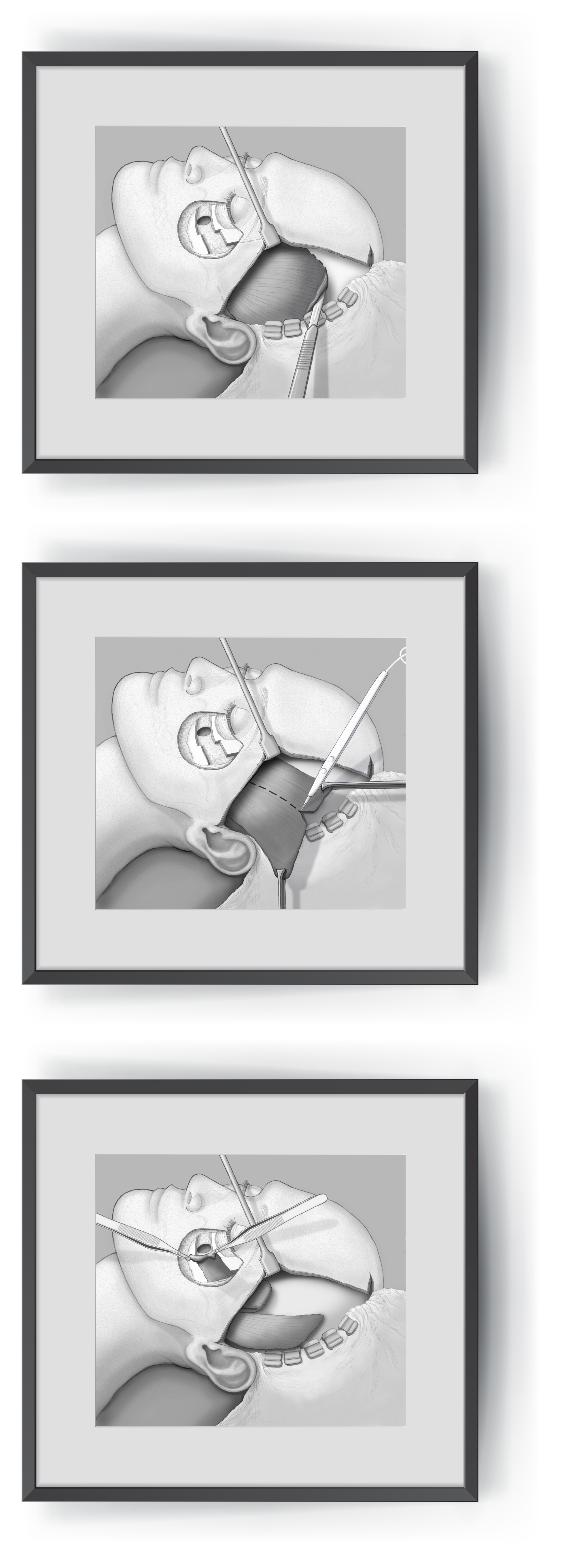
Facial Tumour Removal Surgery
1
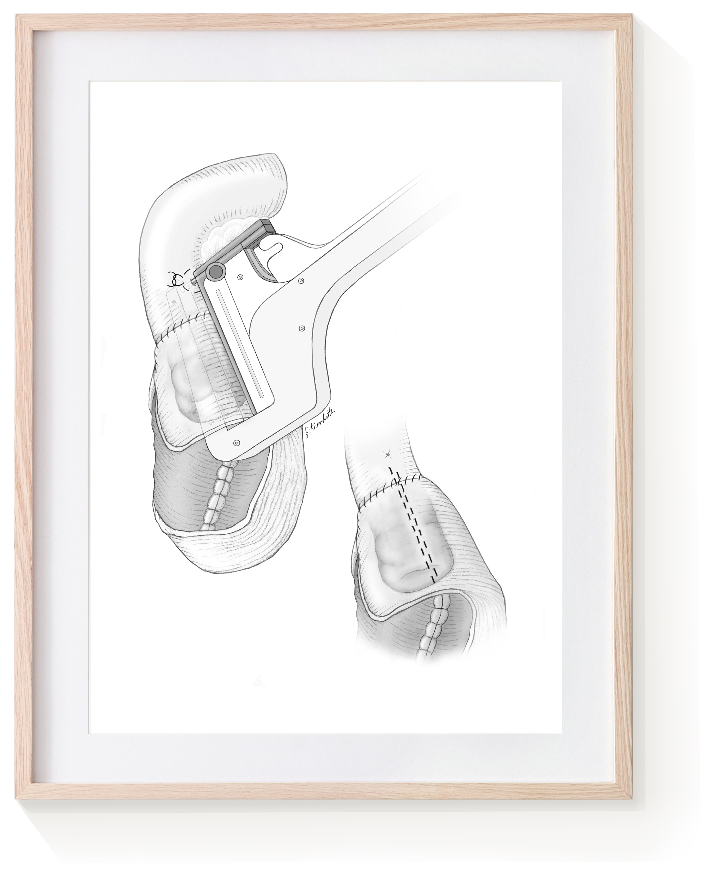
Kock Pouch
1
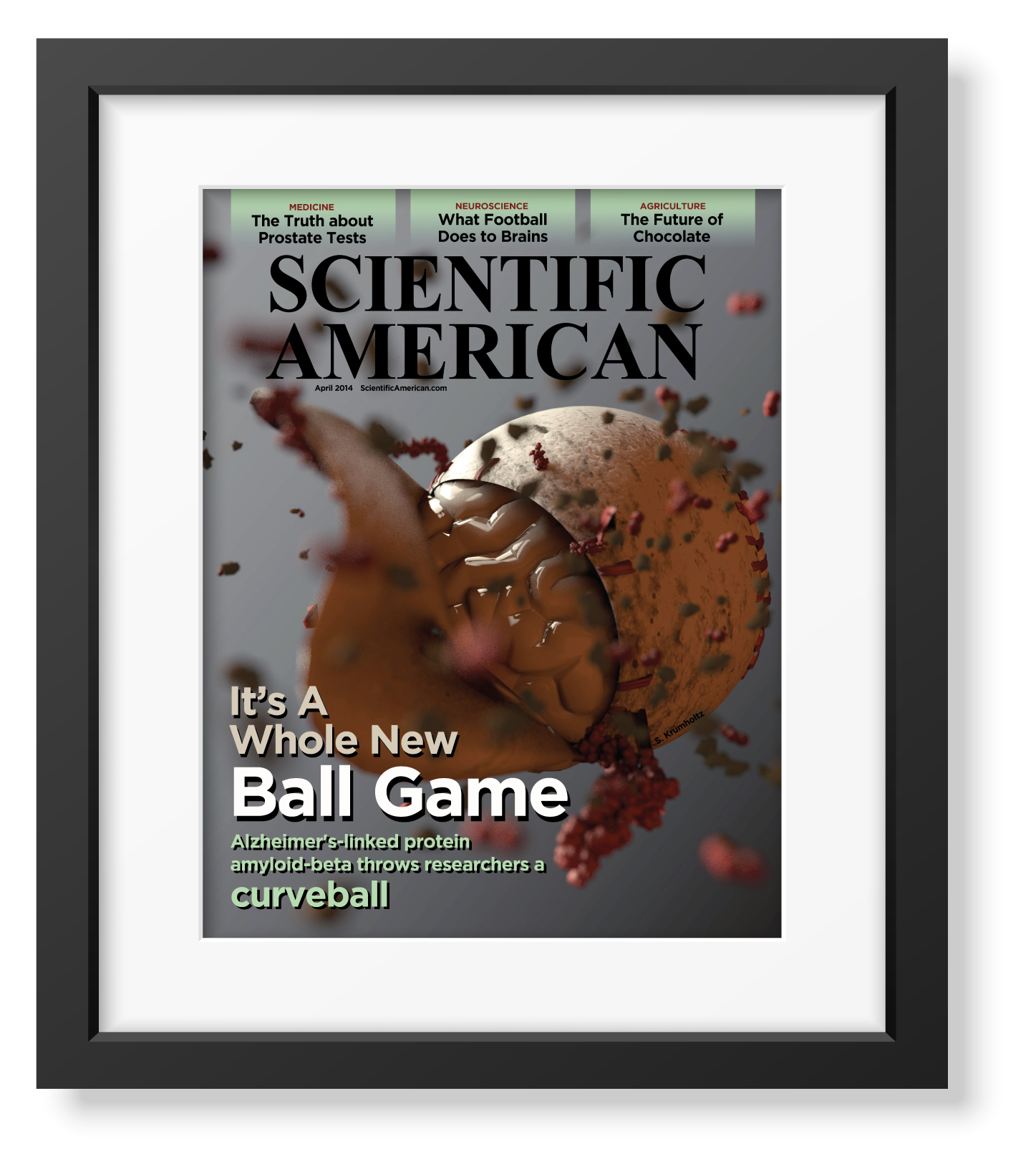
Alzheimer's Mock Cover
2
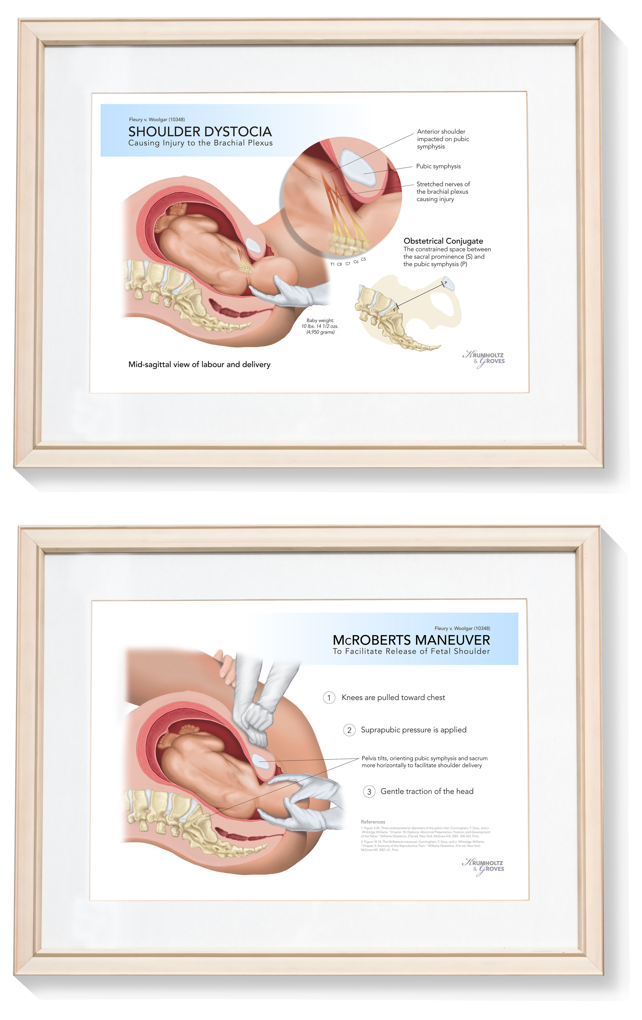
Shoulder Dystocia
2
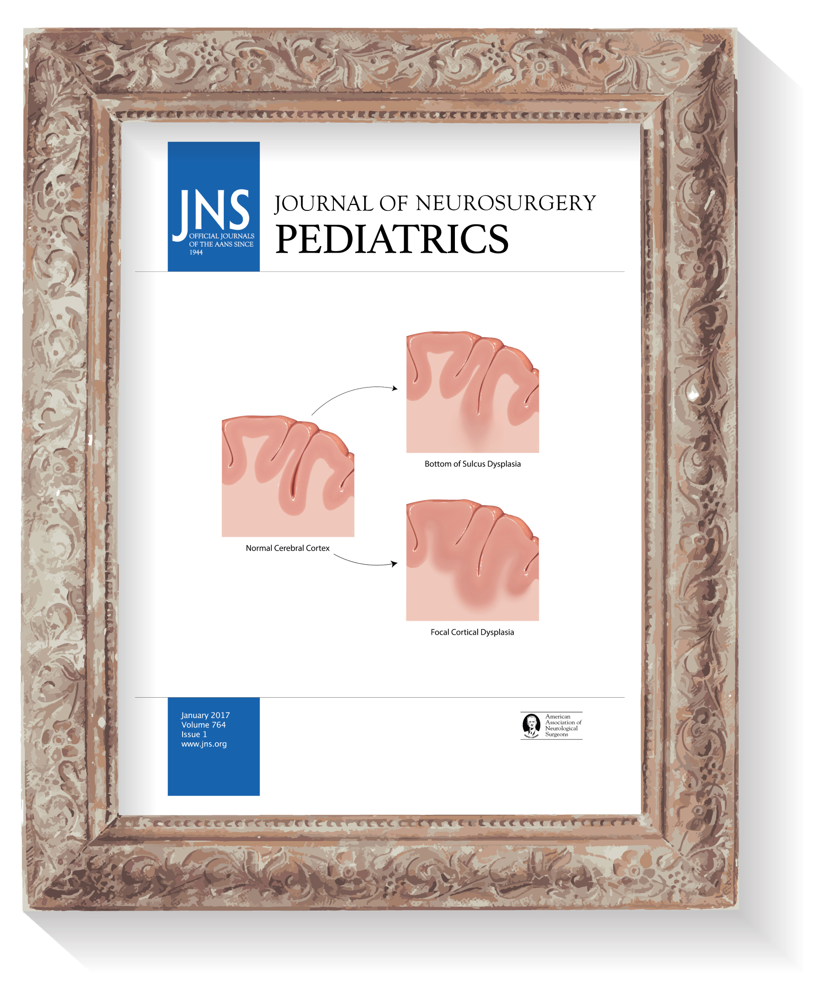
Dysplasia
1
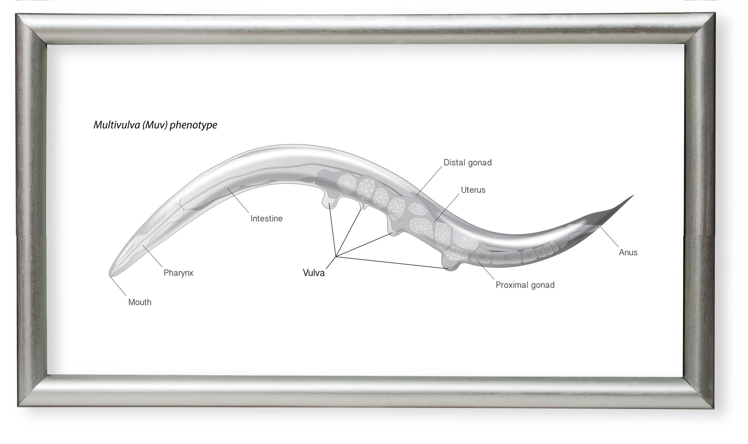
C. elegans
1
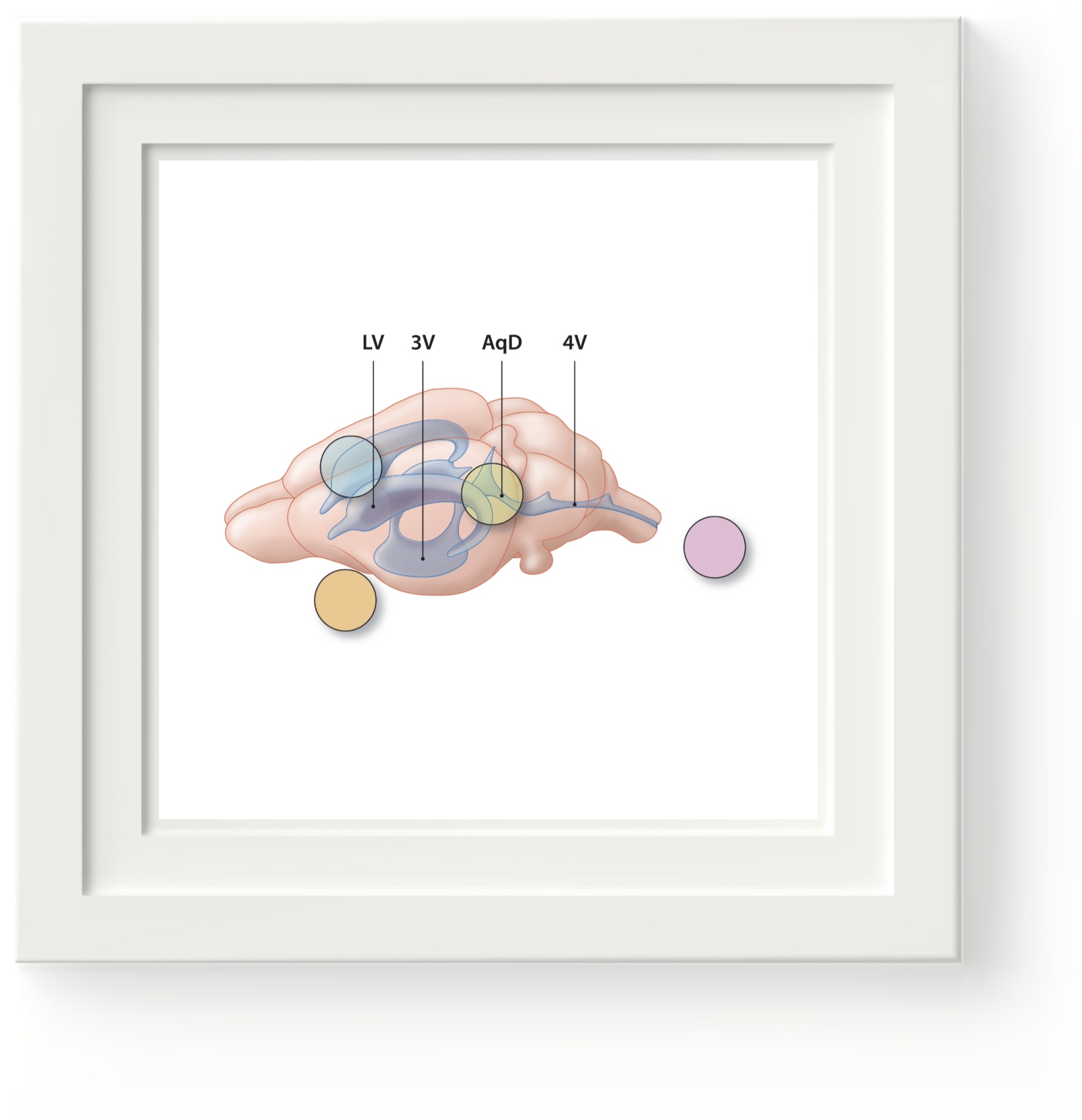
Mouse Brain
2
VP Shunt Part I
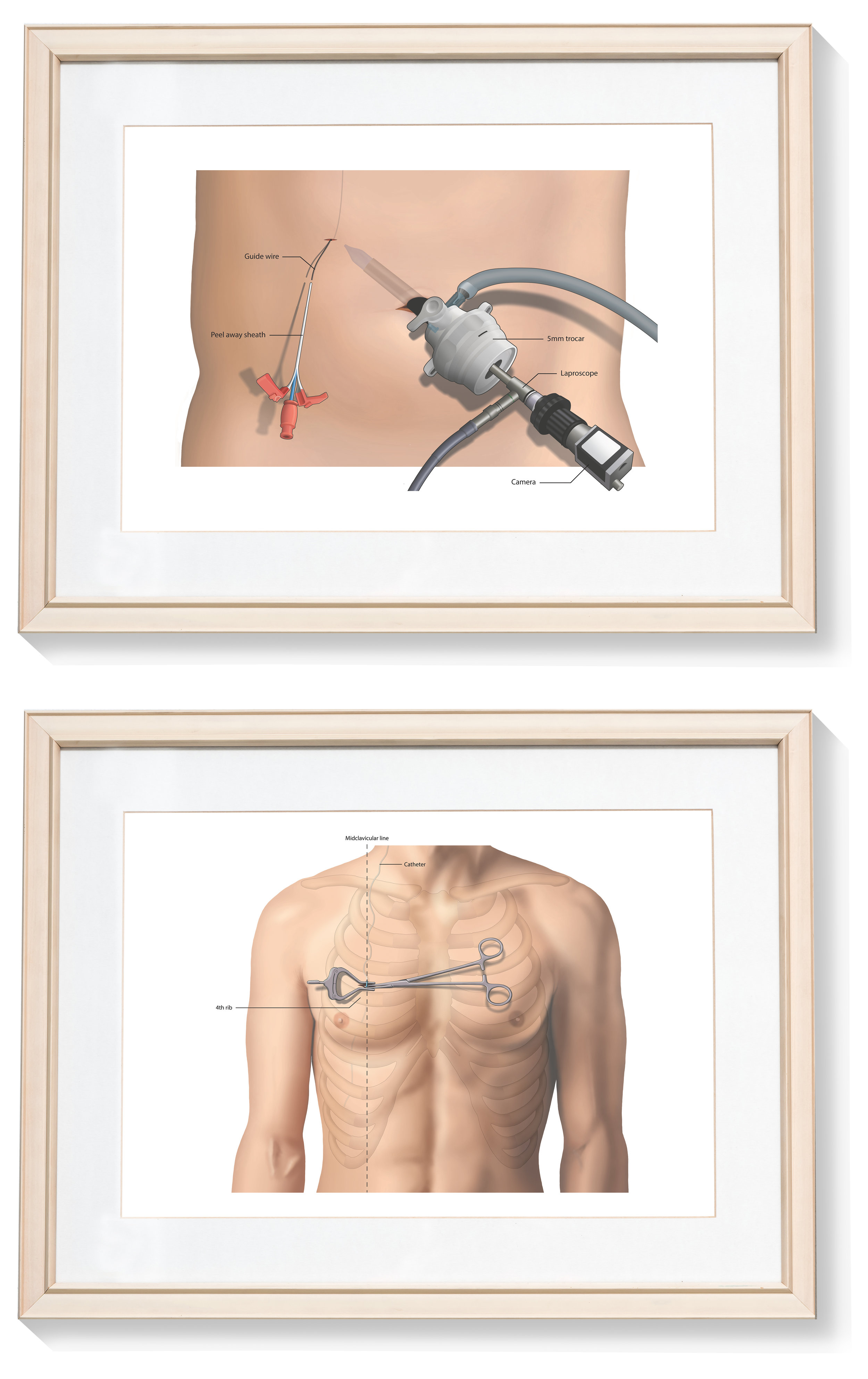
2
VP Shunt Part II
3
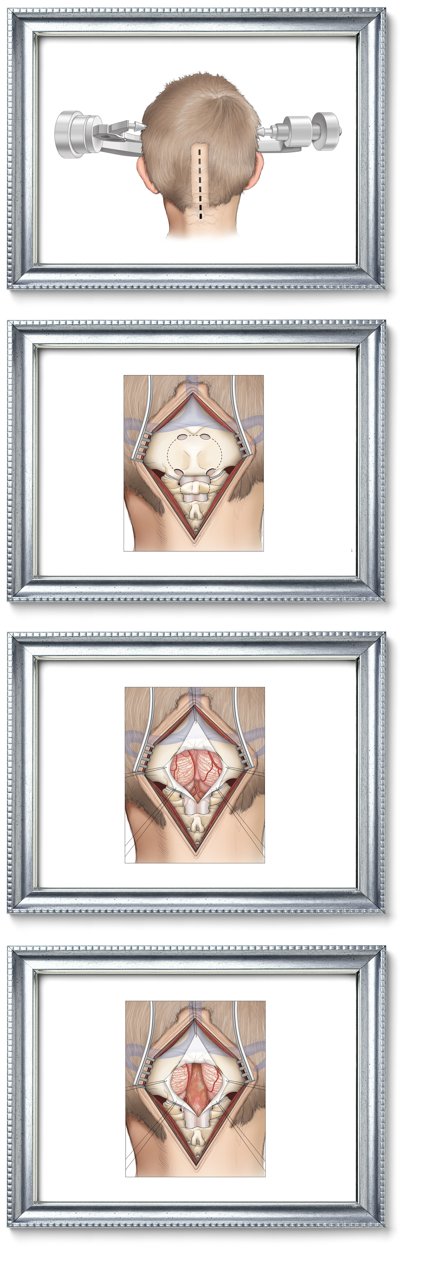
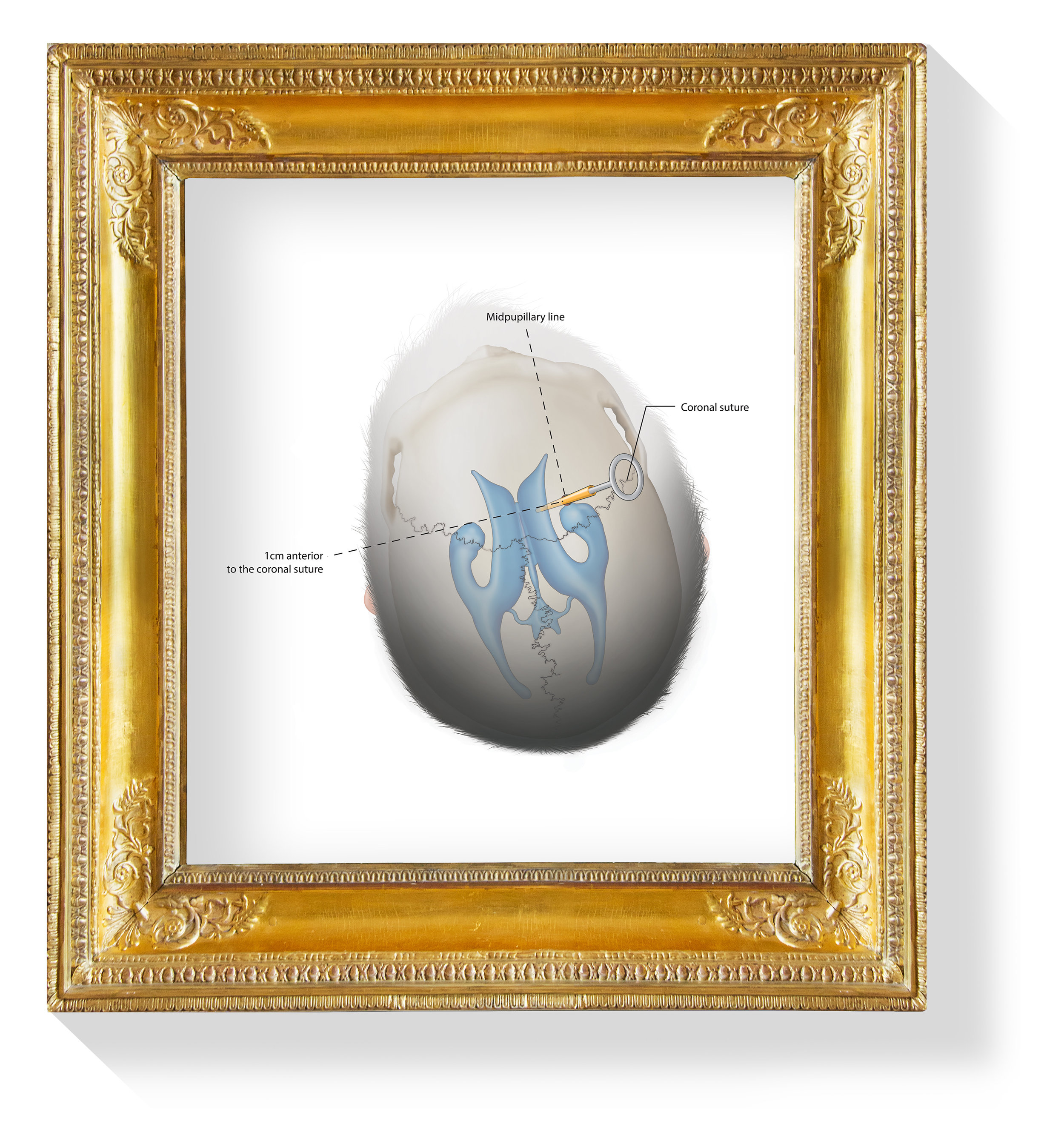
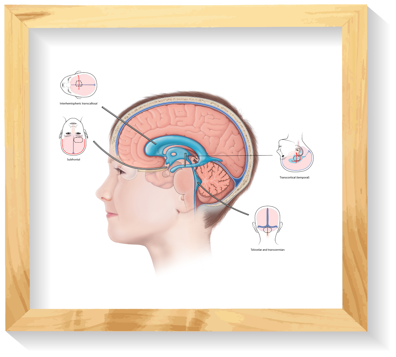
Surgical Approaches to the Ventricular System
1
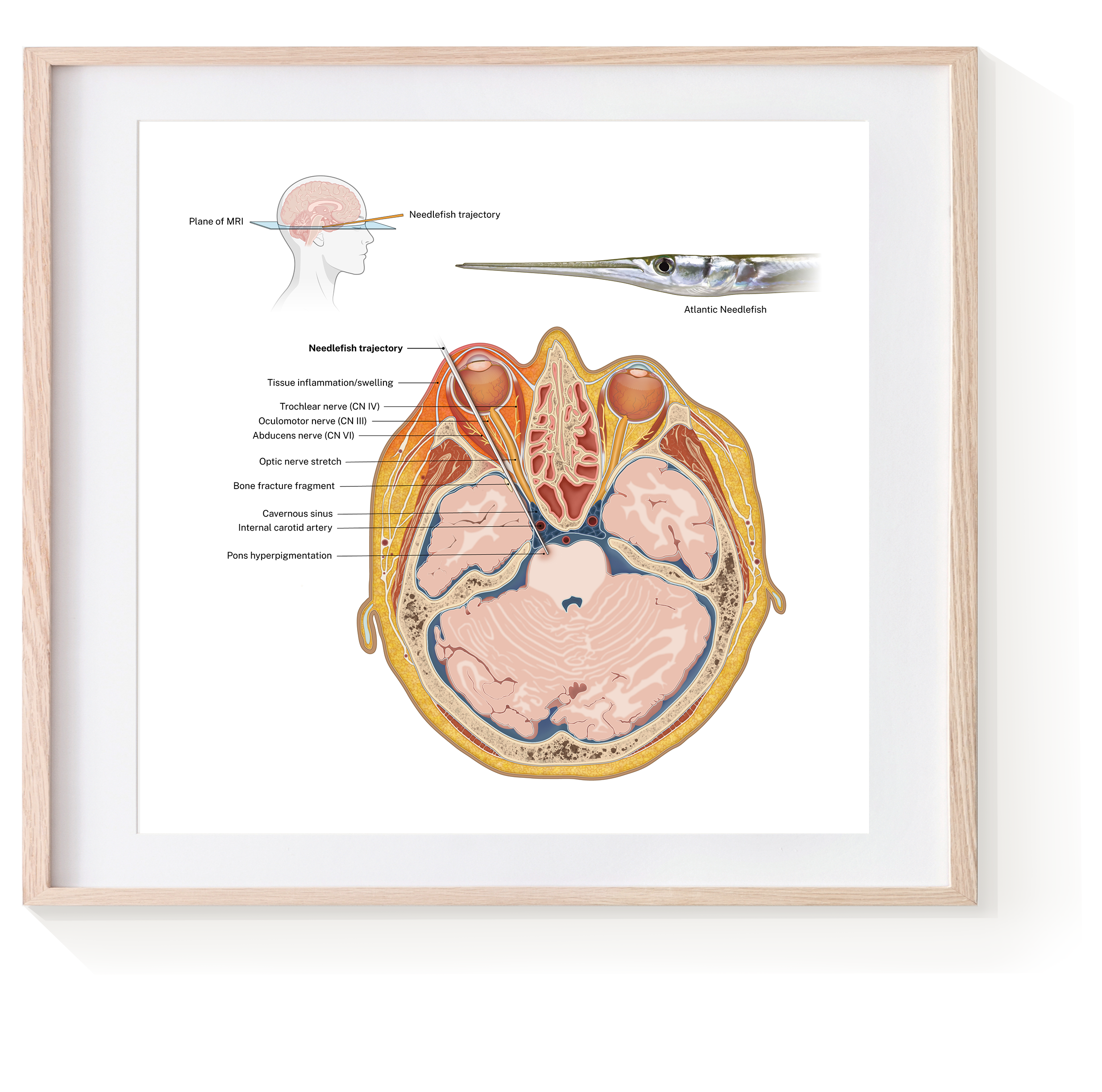
Needlefish Injury Case